
Ultrasound for Endometriosis
“Diagnosis is Treatment”
Diagnosis of Endometriosis with Transvaginal Ultrasound (TVUS) Laparoscopy has, for a long time, been considered the gold standard of diagnosing endometriosis. Since the early 2000s, there has been a significant improvement in the diagnosis, staging, and ability to plan surgical intervention – through using the technique of transvaginal ultrasound (TVUS). It is important to note straight away that a negative finding with TVUS does not necessarily mean that you don’t have endometriosis; an ultrasound performed through the abdominal wall is not accurate enough to rule out pelvic endometriosis.
Dr Wynn-Williams has extensive experience in performing a TVUS on deep infiltrating endometriosis (DIE). Resent research has shown that having the capacity to perform the diagnostic endometriosis ultrasound during the patient examination process significantly enhanced the patients’ experience. Endometriosis TVUS enables Dr Wynn-Williams to plan the extent of often very complex pelvic surgery..

Deep Infiltrating Endometriosis (DIE)
Endometriosis tissue will typically grow in areas deep in the pelvis, such as behind the uterus, between the vagina and rectum (Pouch of Douglas), in the rectum, the ovaries, and the bladder. Deep infiltrating endometriosis is the definition given to endometriosis that has invaded at least 5mm deep into the tissue. Often, it is only the most experienced practitioners who can spot these areas of deep endometriosis when they position the ultrasound probe up against the diseased regions, through the back of the vagina. The areas of endometriosis appear as darker patches as they are denser with the scar tissue they contain. Because Dr Wynn-Williams performs surgery and excises endometriosis without delay - often only a few days after he has completed the ultrasound scan - he is continuously improving his skill in recognising lesions of deep infiltrating endometriosis.
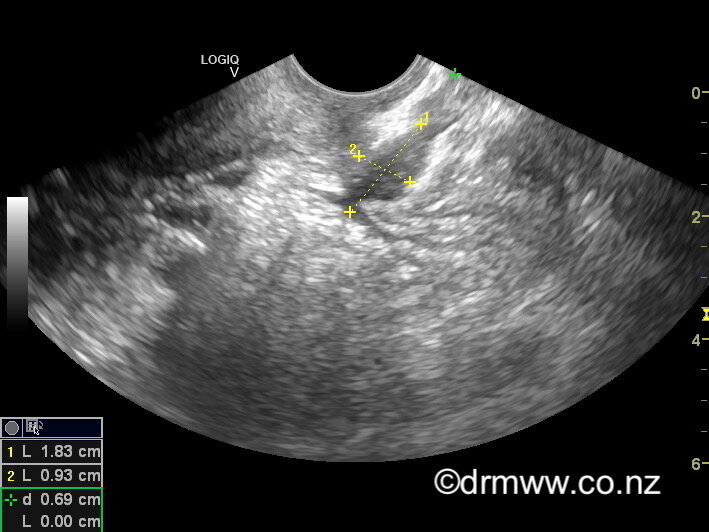
A rectal muscularis endometriosis lesion seen on TVUS
Bowel Endometriosis
Endometriosis tissue that grows in the bowel (rectum, sigmoid, and sometimes the caecum) will often cause symptoms such as painful bowel motions (dyschezia), rectal bleeding, constipation, diarrhoea, and long skinny bowel motions (ribbon stools). In practised hands, bowel lesions can be seen quite clearly using TVUS. The ultrasound allows Dr Wynn-Williams to work out the size and number of bowel lesions present and give you several options for how to manage your symptoms. If you have nodules in the bowel that are larger than 3cm or multiple areas of bowel endometriosis, you may be referred to a colorectal surgeon as part of your management plan. The majority of lesions will be smaller than 3cm and will be managed by Dr Wynn-Williams himself. If the extent of the endometriosis in the bowel or bladder is seen to be extensive, then you may be referred for an MRI scan. Endometriosis on the small intestine cannot be seen on TVUS.

A rectal muscularis lesion after laparoscopic shaving
A “Negative” Endometriosis Ultrasound
TVUS can how the extent of pelvic adhesions (organs stuck together) that may have been caused by endometriosis. It can also help identify the size and number of endometriosis cysts (endometriomas) or other potential pathology that could be affecting the ovaries.
Pelvic ultrasound will generally not pick up the superficial endometriosis that the majority of women with endometriosis have, and that is why a laparoscopy may still be required despite a negative ultrasound. Often, pelvic ultrasounds are performed by sonographers and radiologists who do not have experience in finding and locating deep infiltrating endometriosis. If you have symptoms and are concerned about endometriosis, it is essential that your pelvic ultrasound is performed by someone with extensive experience in the field.
Dr Wynn-Williams will work with you to diagnose and treat your endometriosis and pelvic pain, using the latest in advanced ultrasound and surgical techniques.

Areas on DIE that can be seen on TVUS